Through Our Eyes
A Gallery of Personal Visual Perceptions
Contributed by the Macular Degeneration Community
The following visual field photos were created from information submitted by the MD Community.
 |
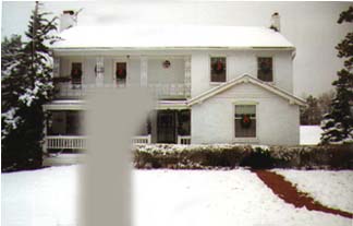
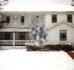
Subject: Dan Roberts. Pictured: Left eye. Map of scotoma derived from computerized perimetry test, compliments of Nidek Technologies, Inc. Condition: central serous chorioretinopathy. Right eye in early stages. |
 |
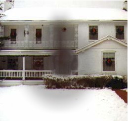
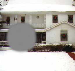
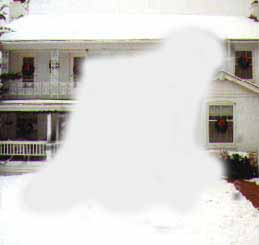
Subject: Donna Broadstock. Pictured: right eye only. Condition: wet form, macular degeneration. Early stages of dry form in left eye. |
 |
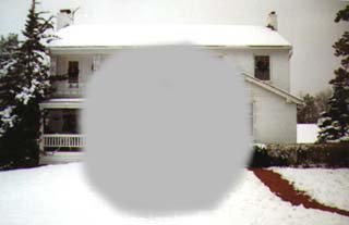
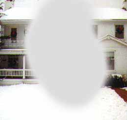
Subject: Bob Thompson. Perception shown: both eyes. Condition: cone-rod dystrophy (best diagnosis to date). |
 |
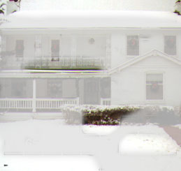
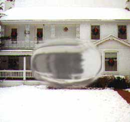
Subject: Lara. Pictured: both eyes. Condition: Stargardt’s syndrome. |
 |
Subject: Joan Micone. Pictured: both eyes. Condition: Stargardt’s macular dystrophy (Fundus Flavimaculatus).Constantly shifting waves of distortion. |
 |
Subject: Colen Benfield. Pictured: both eyes. Condition: wet form, macular degeneration. Right eye in early stages with blind area and surrounding dimness covering most of lower left quadrant of VF Grid. No central vision in left eye except 1st column of grid. Vitrectomy surgery performed on right eye in late 1998. |
 |
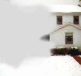
Subject: Linda Olsen. Pictured: left eye only. Condition: Macular Degeneration. |
 |
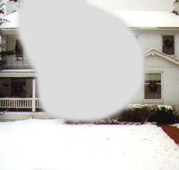
Subject: Pam Moore. Pictured: right eye only. Condition: Macular Degeneration |
 |
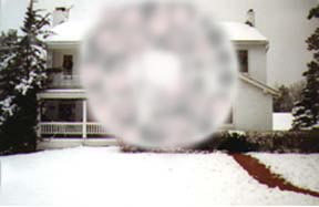
Subject: Barbara Brown. Pictured: both eyes. Condition: Macular Degeneration with bilateral drusen. Diagnosed at age 50 in 1996. |
 |
Subject: Alan Wilson. Pictured: both eyes. Condition: Left eye diagnosed as MD with choroidal neovascularization, Sept. 1999. Right eye rediagnosed as possible serpiginous choroiditis. |
 |
Subject: Bert Schaefer. Pictured: left eye only. Condition: Wet MD. |
 |
Subject: Cheri Matthew. Pictured: both eyes. Condition: Stargardt’s. Visual imaging has been as pictured since age 51 in the year 2000. Reading text is similar to Joan Micone’s example (above) with addition of blind areas. |
 |
Subject: Jody Thomann. Pictured: left eye. Condition: Edema caused by early stage dry macular degeneration. |
 |
Subject: Fred Hemmer. Pictured: both eyes. Condition: Macular degeneration. |
To see through the eyes of our RP friends, visit Through Our Eyes II