Anatomy of the Eye
by Dan Roberts
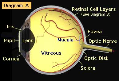
Cornea: The transparent part of the eyeball which covers the iris and pupil. A tear film normally coats the cornea, keeping the eye moist. Directly behind the cornea is the aqueous chamber, which is filled with a clear fluid for the purpose of maintaining the pressure of the eye.
Iris: The colored diaphragm in the anterior chamber of the eyeball which contracts and expands to adjust for light intensity.
Pupil: The opening in the center of the iris through which light passes.
Lens: The transparent, dual-convex body which focuses light rays onto the retina. It is normally capable of changing shape to allow the eye to focus on both near and distant images.
Retinal Cell Layers: The membrane on the inner wall of the eyeball which receives the image from the lens and converts it into nerve impulses.
Vitreous Gel: A clear jelly-like substance which fills the posterior chamber of the eyeball. Normally attached to the retina, it can become detached in clumps or strings, called “floaters,” which are usually harmless, but can cast annoying shadows on the retina.
Fovea: The center of the retina. The region of highest visual acuity and cone cell density.
Macula: The area in and around the fovea. Made up mostly of cone cells, it is responsible for central vision and contains the fovea. The macula is less than 5% of the total retina.
Sclera: The white, dense, fibrous outer coating of the eyeball.
Optic nerve: Transmits nerve impulses from the retinal cell layers to the brain.
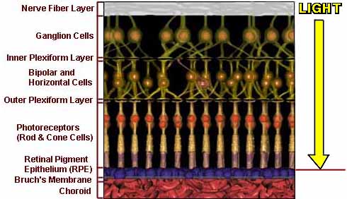
Light first enters the optic (or nerve) fiber layer and the ganglion cell layer, under which most of the nourishing blood vessels of the retina are located. This is where the nerves begin, picking up the impulses from the retina and transmitting them to the brain.
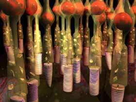
Light is transmitted through the several neural layers by Müller glial cells that act as optic fibers. The light is received by photoreceptor cells called rods (responsible for peripheral and dim light vision) and cones (providing central, bright light, fine detail, and color vision). The photoreceptors convert light into nerve impulses, which are then processed by the retina and sent through nerve fibers to the brain. The nerve fibers exit the eyeball at the optic disk and reach the brain through the optic nerve.
Directly beneath the photoreceptor cells is a single layer of retinal pigment epithelium (RPE) cells, which nourish the photoreceptors. These cells are fed by the blood vessels in the choroid. The condition called wet (or exudative) macular degeneration results when new vessels grow from the choroid through Bruch’s membrane, and into spaces above and below the RPE. These new vessels, or neovascularization, are fragile, and they often bleed. If not stopped in time, bleeding can permanently damage the photoreceptors, replacing them with non-seeing scar tissue.
Photoreceptors can also wither as a result of age, causing dry (or atrophic) macular degeneration. This condition (also called age-related macular degeneration, or ARMD) is usually less severe than wet macular degeneration, with central vision diminishing over a period of years.
Illustration of the Retina:
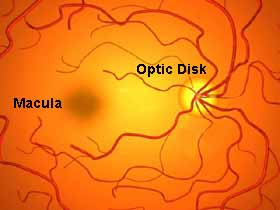
This is a view of the retina at the back of the eyeball. The retina, which lines the inside of the eyeball, is essentially transparent, so a doctor can see through all of the layers to the choroid. The choroid is where the blood vessels are located, which give the retina it’s reddish hue. This also explains why “red eye” often occurs in photographs.
The retina captures light like the film in a camera. Film, however, is different, in that it is just as sensitive at its edge as in its center. Only the center of the retina, the macula, can see fine detail.
For more information about the various types of macular degeneration, see “What Is Macular Degeneration?.”
View an animated depiction of the individual parts of the eye.
|
© Dan Roberts, 2001
The content of this page has been reviewed for accuracy by Martin A. Mainster, Ph.D., M.D., FRCOphth. Art work by Evayla Computer Graphics
Kansas City, Missouri |